|
|
TshFNA-Examiner: A Nuclei Segmentation and Cancer Assessment Framework for Thyroid Cytology Image
KE Jing1(柯晶), ZHU Junchao2 (朱俊超), YANG Xin1(杨鑫), ZHANG Haolin3 (张浩林), SUN Yuxiang1(孙宇翔), WANG Jiayi1(王嘉怡), LU Yizhou4(鲁亦舟), SHEN Yiqing5(沈逸卿), LIU Sheng6(刘晟), JIANG Fusong7(蒋伏松), HUANG Qin8(黄琴)
2024, 29 (6):
945-957.
doi: 10.1007/s12204-024-2743-y
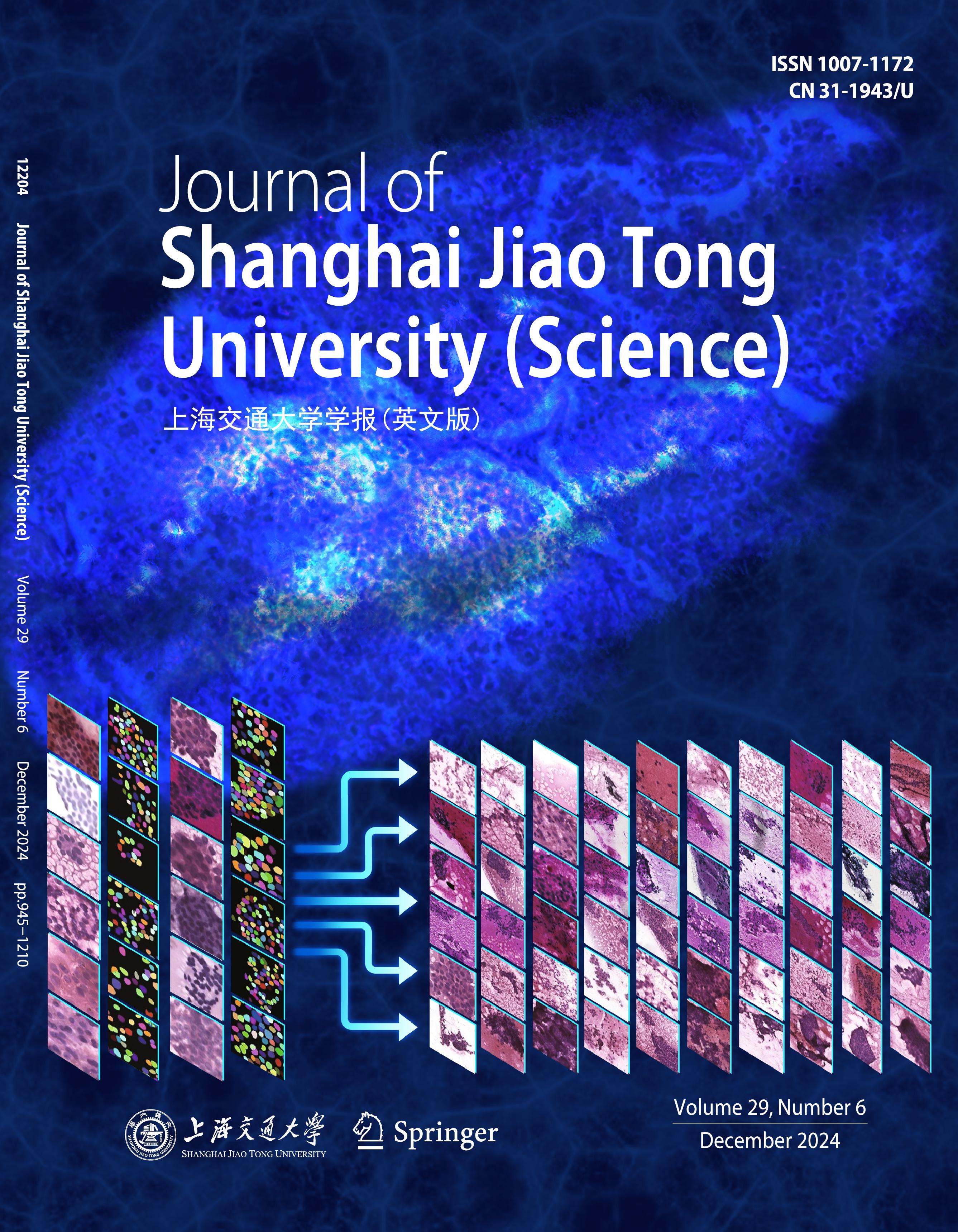
Examining thyroid fine-needle aspiration (FNA) can grade cancer risks, derive prognostic informa�tion, and guide follow-up care or surgery. The digitization of biopsy and deep learning techniques has recently enabled computational pathology. However, there is still lack of systematic diagnostic system for the complicated gigapixel cytopathology images, which can match physician-level basic perception. In this study, we design a deep learning framework, thyroid segmentation and hierarchy fine-needle aspiration (TshFNA)-Examiner to quantita�tively profile the cancer risk of a thyroid FNA image. In the TshFNA-Examiner, cellular-intensive areas strongly correlated with diagnostic medical information are detected by a nuclei segmentation neural network; cell-level image patches are catalogued following The Bethesda System for Reporting Thyroid Cytopathology (TBSRTC) system, by a classification neural network which is further enhanced by leveraging unlabeled data. A cohort of 333 thyroid FNA cases collected from 2019 to 2022 from I to VI is studied, with pixel-wise and image-wise image patches annotated. Empirically, TshFNA-Examiner is evaluated with comprehensive metrics and multiple tasks to demonstrate its superiority to state-of-the-art deep learning approaches. The average performance of cellular area segmentation achieves a Dice of 0.931 and Jaccard index of 0.871. The cancer risk classifier achieves a macro-F1-score of 0.959, macro-AUC of 0.998, and accuracy of 0.959 following TBSRTC. The corresponding metrics can be enhanced to a macro-F1-score of 0.970, macro-AUC of 0.999, and accuracy of 0.970 by leveraging informative unlabeled data. In clinical practice, TshFNA-Examiner can help cytologists to visualize the output of deep learning networks in a convenient way to facilitate making the final decision.
References |
Related Articles |
Metrics
|

