

目前KOA传统治疗方法包括物理治疗、药物治疗、手术治疗及膝关节矫形器治疗等,相比创伤较大的手术治疗、物理治疗或药物治疗等方式[3],膝关节矫形器通常是依靠三(四)点力学原理矫正膝关节的异常下肢力线.本文采用了上海第九人民医院王金武团队研发的增材制造(AM)膝关节矫形器,AM俗称3D打印,是通过数字控制利用逐层堆积的方式制造物体的技术.随着计算机技术的发展,3D打印在个性化矫形器的应用日益广泛.AM膝关节矫形器在减轻支具重量、提升佩戴舒适度的同时,通过对膝关节两侧分别施加不同大小的轴向拉伸力,矫正力线,减轻患者患侧负荷,对对侧局部减荷,提高临床治疗效果,给患者带来良好的康复体验.
1 实验材料与方法
1.1 实验材料
硬件设备为飞利浦PHILIPS Brilliance 64排螺旋CT机(飞利浦公司,荷兰),由上海交通大学第九人民医院影像科提供;Structure Sensor便携式扫描仪(Occipital,美国).
软件设备为Mimics21.0(Materialise公司,比利时);CATIA P3 V5-6R2020(达索公司,法国);Geomagic Wrap 2021( 3D Systems公司, 美国);Ansys Workbench 2020 R2(ANSYS公司,美国);MATLAB R2021b(MathWorks公司,美国).
1.2 实验对象
选择一名患有KOA的女性志愿者,年龄71岁,体重60 kg,使用CT机对实验对象膝关节进行扫描,在扫描成像过程中使其膝关节处于非负重位,足趾垂直于水平面,保持髌骨在上,最终得到层厚为0.625 mm、大小为512像素×512像素的下肢全长图像.实验对象在试验前充分了解试验可能带来的风险并签署知情同意书,且获得上海交通大学医学院附属第九人民医院医学伦理委员会的批准.
1.3 实验过程
1.3.1 建立膝关节-矫形器整体三维模型
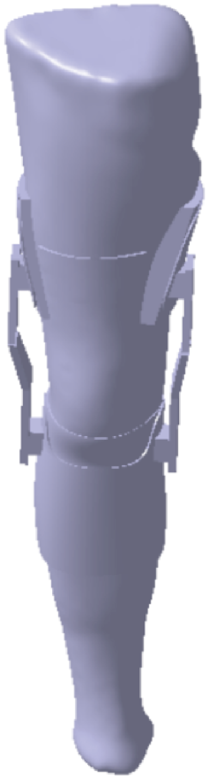
将膝关节CT数据导入Mimics,利用蒙板工具、灰度测量、阈值选择、区域增长等操作逐步提取出膝关节股骨、胫骨、腓骨、髌骨的轮廓.三维重建膝关节各部分几何模型后,将其以STL格式导入 Geomagic Wrap 软件进行精确曲面处理,结合三维快速扫描仪获得患者下肢患侧膝关节体表点云数据,通过编辑轮廓线、构造曲面片、拟合曲面等操作分别对各部件点云数据进行加工处理,完成模型修整及曲面生成[8],通过参数交换将STL格式转为三维图形文件的STP格式导入Catia,并对数据进行三维实体建模.利用三维建模软件Catia进行布尔运算、草图绘制、拉伸等操作,实体构建包含大腿袖、小腿袖、软垫、绑带和关节铰链等结构的定制式AM膝关节矫形器.由于在CT图像中软组织显示界限不清,故关节软骨和内外侧半月板利用Catia在CT资料的显影基础上基于膝关节解剖特点三维重建形成实体模型[9-10].将各部件装配成型后,重建的膝关节及膝关节-矫形器整体三维模型如图1和图2所示.
图1
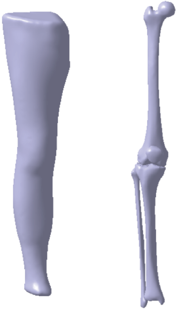
图2
1.3.2 建立膝关节-矫形器整体有限元模型
图3
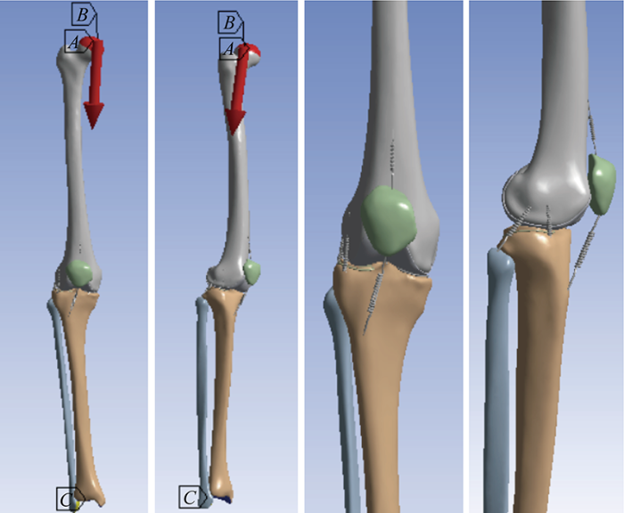
图4
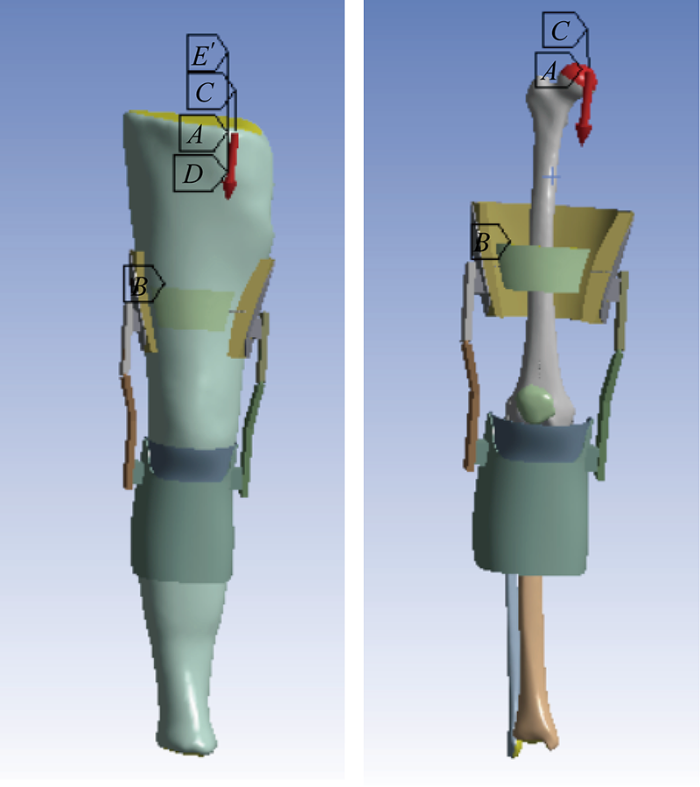
表1 膝周主要韧带刚度系数
Tab.1
| 韧带 | 刚度系数/(N·mm-1) |
|---|---|
| 前外侧副韧带 | 2 144 |
| 后外侧副韧带 | 2 144 |
| 内侧副韧带 | 134 |
| 前交叉韧带 | 201 |
| 后交叉韧带 | 258 |
| 髌骨韧带 | 300 |
1.3.3 材料属性
表2 骨骼、软骨、半月板及矫形器的材料属性
Tab.2
| 组织结构 | 弹性模量E/MPa | 泊松比ν |
|---|---|---|
| 骨骼 | 12 000 | 0.3 |
| 软骨 | 5 | 0.46 |
| 半月板 | 50 | 0.45 |
| 壳体及关节铰链 | 1 000 | 0.2 |
| 软垫 | 50 | 0.45 |
1.3.4 边界、加载条件和网格划分
使用Workbench软件设置膝关节各结构的相互作用、边界条件以及载荷,根据实验加载条件设置以下接触区.
膝关节:软组织与胫骨、腓骨、股骨、髌骨、股骨软骨间各设1个接触对,内外侧胫骨软骨与内外侧半月板间设2个接触对,股骨软骨与内外侧胫骨软骨间设2个接触对,胫骨与腓骨间设2个接触对,胫骨与胫骨软骨间设2个接触对,股骨软骨与股骨间设1个接触对,共14对,均定义为绑定接触;股骨软骨与内、外侧半月板、胫骨及髌骨,共4对,定义均为无摩擦接触.由于对称接触观察接触面的压力值不是正确压力值,非对称接触观察接触面的压力值为真实接触面压力值,故除胫腓骨(2对)为对称行为外,其他接触行为皆为非对称行为.
矫形器:大腿壳体与上软垫及上支关节铰链、软组织与软垫、小腿壳体与下软垫及下支关节铰链(共8对),均定义为绑定接触;上下支关节铰链、软组织与绑带(共4对),定义均为无摩擦接触.除上下支关节铰链(2对)为对称行为外,其他接触行为皆为非对称行为.
股骨的6个自由度不受约束,固定胫腓骨,将边界条件设置在胫骨远端和腓骨远端,固定6个自由度.赵春霞等[16]研究表明,在矫形器内衬材料一定的情况下,矫形器的上铰链受到的压力越大, 矫形器的减荷能力越大.故在膝关节矫形器模型患侧的铰链连接部位施加-100 N的轴向载荷,在健侧施加 -10 N 的轴向载荷,以此模拟矫形器撑开膝关节的矫正作用.根据Park等[17]研究,在股骨近端沿下肢负重轴方向施加 1 100N (约2倍重力)的压缩载荷,以此对比佩戴定制式增材制造膝关节矫形器前后的膝关节生物力学变化.最后对膝关节及矫形器进行网格划分,单元类型为二阶四面体单元,具体单元数和节点数如表3所示.根据文献[7,18]可知,目前大多数膝关节有限元研究中模型未经过网格收敛测试,有关此类有限元分析常利用对比其他文献中实验数据的方法来代替网格收敛性检查,本文模型大部分部件单元数和节点数已超越张刘会等[19]研究内模型各部件数目,且网格更为精细,故可以证明本实验网格收敛性.
表3 膝关节-矫形器整体模型的各部件单元数和节点数
Tab.3
| 各部位 网格划分 | 单元数 | 节点数 | 各部位 网格划分 | 单元数 | 节点数 |
|---|---|---|---|---|---|
| 髌骨 | 1 483 | 2 439 | 股骨软骨 | 48 648 | 90 410 |
| 胫骨 | 24 122 | 36 463 | 半月板 | 9 803 | 17 432 |
| 股骨 | 42 404 | 63 464 | 软垫 | 135 631 | 222 446 |
| 腓骨 | 5 616 | 9 785 | 壳体 | 82 636 | 152 147 |
| 胫骨软骨 | 20 437 | 36 602 | 关节铰链 | 48 718 | 79 022 |
2 结果
2.1 膝关节Von Mises应力和位移
Von Mises应力[20]是一种等效应力,是根据第四强度理论得到的当量应力,为综合概念,考虑了第一、第二和第三主应力,可以对疲劳、破坏等进行评价.Von Mises应力可以清晰描述出一种结果在整个模型中的变化,从而使研究者快速确定模型中的最危险区域.Von Mises应力根据3个方向的主应力计算得到,计算公式为
式中:σ1,σ2,σ3分别为第一、第二和第三主应力.本实验采用Von Mises应力对比佩戴定制式AM膝关节矫形器前后的膝关节生物力学变化.
对膝关节施加2倍重力沿下肢负重轴方向的压缩载荷作用, 两种情况下膝关节的Von Mises应力分布如图5和图6所示.其中,A1~E1分别为股骨软骨,内、外侧半月板,内、外侧胫骨软骨,A2~E2分别为股骨软骨,内、外侧半月板,内、外侧胫骨软骨.未佩戴矫形器和佩戴矫形器两种情况下股骨软骨,内、外侧半月板和内、外侧胫骨软骨最大Von Mises 应力分别为 0.62与0.14、0.98与0.55、0.007 6 与 0.054 1、0.44与 0.14、0.000 3 与 0.012 3 MPa.其各部位软骨、内外侧半月板分布最大Von Mises应力值分布的整体情况与文献[9,21]中有限元分析结果较为接近.分布上,未佩戴矫形器情况下股骨软骨最大Von Mises应力分布于股骨后侧及与半月板接触位置,内侧半月板主要分布于中部以及前后角位置,外侧半月板主要分布在前角位置,胫骨软骨应力主要分布于内部的中心及与半月板接触位置.佩戴矫形器情况下股骨软骨最大Von Mises应力主要分布于内侧与半月板交接部位,其余部位最大应力分布明显由内侧向外侧转移,达到较平衡状态.位移方面,佩戴矫形器前后情况下膝关节股骨相对于胫骨前向上的位移分别为13.96和0.59 mm,股骨前移状态有明显改善,关节间的稳定性增强.
图5
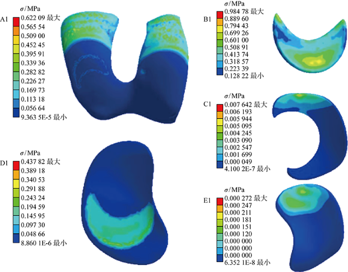
图5
未佩戴矫形器时股骨软骨、内外侧半月板、内外侧胫骨软骨的VonMises应力云图
Fig.5
VonMises stress cloud map of femoral cartilage, medial and lateral meniscus, medial and lateral tibial cartilage without orthosis
图6
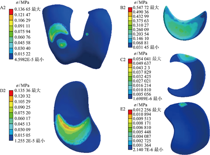
图6
佩戴矫形器时股骨软骨、内外侧半月板、内外侧胫骨软骨的VonMises应力云图
Fig.6
VonMises stress cloud map of femoral cartilage, medial and lateral meniscus, medial and lateral tibial cartilage when wearing orthosis
2.2 两种情况下的膝关节内翻角度
图7
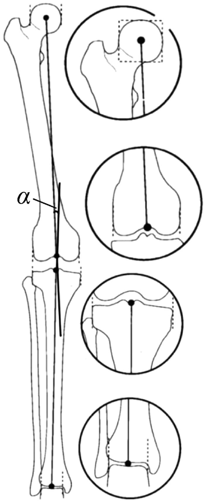
记录4个中心点的具体坐标,在膝关节有限元模型中分别通过对股骨、胫骨进行X、Y、Z轴的定向位移,评估所有结果后,将位移结果导入MATLAB,利用克莱姆法则得到4个中心点邻近坐标,通过反三角函数运算得出轴间夹角,测出佩戴矫形器前后情况下膝关节内翻倾角分别为7.84°和6.65°.
3 讨论
3.1 定制式AM膝关节矫形器治疗优势
常见KOA的治疗方式包括物理治疗、药物治疗以及手术治疗.物理治疗及药物治疗虽可以一定程度减轻患者的疼痛,但不能改变患者步行中异常的膝关节受力方式,即未矫正下肢力线的偏移,故治疗效果不甚理想;手术治疗主要针对中晚期KOA患者[26],但创伤较大且花费昂贵难以被广泛接受.目前,膝关节矫形器作为一种新型的治疗方式因其无创伤、能有效减轻疼痛且花费较低被广泛运用于早、中期KOA的治疗.膝关节矫形器可有效改善临床症状并提高膝关节的稳定性,对常规治疗无效及因各种原因不能接受手术治疗的KOA患者也有较好疗效[27-28].常见膝关节矫形器包括三(四)点力式与整体免荷式.三(四)点力式膝关节矫形器依靠三(四)点力学受力原理平衡部分膝关节力矩,降低单侧软骨压力,矫正下肢力线,减轻疼痛.这种免荷方式增大患侧髁间隙,减小患侧间室负荷,但同时也增加了对侧髁的负荷,对侧腔室的间隙减小,对侧软骨承受的压力增大,软骨磨损增大.整体免荷式膝关节矫形器通过矫形器分担部分股骨对胫骨的压力,通过增大整个膝关节间室间隙,减小膝关节的整体负荷和关节软骨及骨的摩擦.该类矫形器虽然减少膝关节负荷,却未能从根本上矫正下肢力线.
传统膝关节矫形器通过石膏取模,低温热塑板材等方式进行制作,存在无力学仿真、贴合度差、不透气、不美观等问题,导致无法达到预期的康复效果,产品弃用率高.AM技术的不断发展和研究应用,逐步解决了这个问题.相较于传统康复器具,AM技术能够实现符合人体工程学、结构强度最优化及临床适应性设计的个性化要求,以提高临床矫形效果.
本文采用的定制式AM膝关节矫形器在患者的三维快速扫描模型基础上利用3D打印这种数字化三维重建及快速成型的“自下而上”技术制作成型,其治疗KOA的主要原理综合了三(四)点力式原理和整体免荷式矫形原理的特点,基于整体免荷式矫形原理提出了改进的单侧减荷式矫形原理.通过对膝关节两侧分别施加大小不等的轴向拉伸力,根据作用力与反作用力原理、杠杆平衡原理及力的分解原理,产生一个向力较小的侧方向矫正力矩,在矫正力线、减轻患者患侧负荷的同时,对对侧进行局部减荷,从而避免加剧对侧的磨损,且可根据病情通过调节关节铰链进行个性化分档治疗.该矫形器在实现个性化治疗的同时还减轻了矫形支具的质量,可以完全贴合患者皮肤表面,相比传统膝关节矫形器大幅提升了患者佩戴矫形支具时的舒适度.
实验结果表明,KOA患者在佩带膝关节矫形器后疼痛明显减轻,膝关节稳定性明显增加,日常生活能力评分明显提高[29-30].研究表明Cooper等[31]在对膝关节进行相关有限元分析时,只直接截取单个韧带或者骨骼进行研究,没有全方位考虑膝关节的受力分布情况.Haris等[32]只对受试者健侧膝关节进行研究,缺乏对KOA患者患侧膝关节全方位受力分布情况的分析,且以往利用CT图像进行全膝关节建模的研究存在局限性:以往绝大多数膝关节有限元分析仅包含了骨骼、软骨、半月板和韧带等结构,未加入下肢皮肤组织和膝关节矫形器三维模型进行整体建模分析.本文所建模型通过分析矫形器和患者皮肤,尽可能更加真实模拟膝关节矫形器对关节之间的力传递机制,旨在通过对KOA患者在佩带定制式AM膝关节矫形器前后对膝关节的生物力学变化进行有限元分析,针对矫形器治疗效果定量化研究,深入对骨骼、软组织、矫形器的复杂相互作用的理解,进一步规范膝关节矫形器的减荷疗效评价.
3.2 提供膝关节矫形器临床治疗评价的意义
实验结果表明,佩带矫形器后膝关节内翻角及膝关节在2倍重力下的膝关节内侧间室压力均明显减小.实验发现在佩戴膝关节矫形器后患者患侧膝关节内翻角明显降低,由原先的7.84° 降低至6.65°,其标准也接近Matsumoto等[33]和Wang等[34]研究中的正常人体在步行过程中膝关节的内翻角度范围.股骨软骨 Von Mises 应力与Haris等[32]在 1 080 N 载荷下股骨软骨应力为 3.23 MPa,相比减少了 519.2%;半月板Von Mises 应力与Thienkarochanakul等[21] 800 N 载荷下半月板应力为 4.81 MPa,相比减少 878.1%;胫骨软骨 Von Mises 应力与 Thienkarochanakul等[21]在 1 500 N 压缩载荷作用下胫骨软骨应力为 1.53 MPa,相比减少了 347.1%. 由此可见,正常人体与KOA患者的膝关节受力情况存在较大差距,其原因是施加载荷的不同及健患肢不同所致,相对条件下整体 Von Mises应力接近.
佩戴定制式膝关节矫形器后,内侧半月板Von Mises应力较未佩戴时降低55.6%,外侧半月板Von Mises应力增加711%,内侧胫骨软骨Von Mises 应力较未佩戴时降低30.9%,外侧胫骨软骨Von Mises应力增加4 100%,股骨软骨Von Mises应力较未佩戴时降低22.0%.膝关节内侧间室压力部分转移至外侧,使外侧压力明显增加,内侧压力明显降低.目前,上海第九人民医院王金武团队采用定制式AM膝关节矫形器治疗了本研究的实验对象,观察其佩带定制式AM膝关节矫形器后的膝关节运动学及动力学变化,通过步态实验对佩戴矫形器前后的步态数据进行测算、分析,实验数据结果初步证明,患者佩戴矫形器后,支撑相后期(膝关节承重的重要周期)膝关节外翻角增大,膝关节内侧力线向外侧转移,膝关节内侧压力降低,进一步证实了定制式AM膝关节矫形器对早、中期膝骨性关节炎患者膝关节内侧间室减荷的效果.
本实验采用有限元分析方法为内侧间室KOA患者佩带定制式AM膝关节矫形器前后的膝关节生物力学特性变化提供了客观的评价数据,阐明了定制式AM膝关节矫形器在治疗内侧间室KOA中的生物力学作用机制,更客观地反映了该矫形器的生物力学治疗效果,为评估其骨骼肌肉系统中关节的体内力学提供了依据,增强了对其功能和病理状况的理解,有效验证了对膝关节生理病变分析、设计进一步的康复方案和规范膝关节矫形器的减荷疗效评价的可行性.
3.3 偏倚或不足
本研究主要存在以下不足:样本数量单一,仅考虑膝关节主要韧带,未考虑膝关节周围肌肉组织;仅研究了施加垂直压缩载荷情况下佩带矫形器前后膝关节静态有限元模型瞬间的生物力学变化,未能对长期佩带定制式AM膝关节矫形器后膝关节所产生的生物力学变化进行对比;韧带刚度系数存在比较大的个体差异.研究表明万超等[35]在选取不同韧带刚度系数的有限元模拟中发现,除韧带外其他组织内的应力应变分布和大小变化不大,但选取不同的韧带刚度系数依旧会对有限元模拟的结果产生影响,今后在膝关节相应组织力学性能的设置、模型的构建及验证中需足够重视.
本研究重点为提供较新的研究方法进行分析,故后期会进行更多的样本试验,考虑更全面的膝关节动态有限元模型的力学仿真,进一步对比KOA患者长期佩戴矫形器后发生的改变,结合动态高速荧光双平面透视系统进行步态研究,对比评价膝关节矫形器对KOA患者的减荷效果.
4 结语
AM技术现在正以指数级的速度发展[36],其在康复辅具的应用将更加广泛. 对于早、中期的膝骨关节炎患者,定制式AM膝关节矫形器能够矫正下肢力线,减少患侧的内翻角度及间室负荷、改善患处功能并增加膝关节稳定性,为临床研究提供了重要的参考.但仍在样本数量、软组织的塑造、模型构建的差异等方面存在其局限性.尽管存在不足,但随着医疗技术的创新,KOA的有限元分析研究将会更为完善、成熟.本研究可为KOA更加精准化的诊治及膝关节矫形器减荷疗效评价的规范性提供重要参考.
参考文献
超声波检查在膝骨关节炎早期诊断中的应用价值分析
[J].
Application value analysis of ultrasonic examination in early diagnosis of knee osteoarthritis
[J].
MRI and sonography of the knee in acute reactive arthritis
[J].DOI:10.1097/RHU.0000000000001785 URL [本文引用: 1]
Healthcare and scientific treatment of knee osteoarthritis
[J].
Magnetic resonance imaging of human knee joint functionality under variable compressive in-situ loading and axis alignment
[J].DOI:10.1016/j.jmbbm.2020.103890 URL [本文引用: 1]
Comparison of kinematics and contact mechanics in normal knee and total knee replacements: A computational investigation
[J].
DOI:10.1007/s10439-021-02812-0
PMID:34142278
[本文引用: 1]

An objective of total knee replacement (TKR) is to restore the mechanical function of a normal knee. Joint kinematics and contact mechanics performance are two of the primary indices that indicate the success of TKR devices. The aim of this study was to compare the kinematics and contact mechanics of TKR and normal knee joints. An experimentally evaluated finite-element (FE) knee model was developed and used to investigate the performance of four TKR designs (fixed cruciate-retaining (CR), mobile CR, posterior-stabilized (PS), medial pivot design (MP)) and the normal knee joint during a gait cycle. The predicted kinematic results showed that the MP design presented similar kinematics to those of the normal knee joint and did not demonstrate paradoxical motion of the femur. A considerably larger contact area and lower contact pressure were found on the normal knee joint (1315 mm, and 14.8 MPa, respectively) than on the TKRs, which was consistent with the previous in-vivo fluoroscopic investigation. The mobile CR and PS designs exhibited the smallest and greatest contact pressures of the four TKR designs, respectively. The results of the present study help to understand the kinematics and contact mechanics in the TKR during the gait cycle, and provide comprehensive information about the performance of the normal knee joint.
The effects of different repair methods for a posterior root tear of the lateral meniscus on the biomechanics of the knee: A finite element analysis
[J].
DOI:10.1186/s13018-021-02435-0
[本文引用: 1]
To explore the impact of different repair methods for a lateral meniscus posterior root tear on the biomechanics of the knee joint using finite element analysis.
不同屈曲状态下固定轴和移动轴膝关节胫-股关节的生物力学变化
[J].
Biomechanical changes of the tibial-femoral joint of the knee joint with fixed and moving axes under different flexion states
[J].
人体全膝关节精细有限元模型建立及有效性验证
[J].
Establishment and validation of precise finite element model of human total knee joint
[J].
A simulation case study of knee joint compressive stress during the stance phase in severe knee osteoarthritis using finite element method
[J].
DOI:10.3390/medicina57060550
URL
[本文引用: 2]
Background and Objectives: Medial knee osteoarthritis is known to increase the mechanical load on the medial compartment of the knee joint during walking; however, it is not visually understood how much the mechanical load increases nor where in the medial compartment of the knee joint that load is focused. Therefore, we conducted a simulation study to determine the location and amount of the mechanical load in the medial compartment of the knee joint during the stance phase. Materials and Methods: Subject was a patient with right medial knee osteoarthritis. Computed tomography imaging and gait analysis were performed on subject. The CT image of the right knee was calculated using finite element analysis software. Since this software can set the flexion angle arbitrarily while maintaining the nonuniform material properties of the bone region, the model is constructed by matching the knee joint extension image obtained by CT to the loading response phase of gait analysis. The data of muscle exertion tension and vertical ground reaction force were inserted into the knee joint model created from the computed tomography-based finite element method, and the knee joint compressive stress was calculated. Results: With regard to compressive stress, the tibia showed high stress at 4.10 to 5.36 N/mm2. The femur showed high stress at 4.00 to 6.48 N/mm2. The joint compressive stress on the medial compartment of the knee joint was found to concentrate on the edge of the medial tibial condyle in the medial knee osteoarthritis subject. Conclusions: The measurement method of knee joint compressive stress by computed tomography-based finite element method can visually be a reliable method of measuring joint compressive stress in the medial knee osteoarthritis. This reflects the clinical findings because concentration of stress on the medial knee joint was observed at the medial osteophyte.
Model for in-vivo estimation of stiffness of tibiofemoral joint using MR imaging and FEM analysis
[J].
DOI:10.1186/s12967-021-02977-1
PMID:34281578
[本文引用: 1]
Appropriate structural and material properties are essential for finite-element-modeling (FEM). In knee FEM, structural information could extract through 3D-imaging, but the individual subject's tissue material properties are inaccessible.The current study's purpose was to develop a methodology to estimate the subject-specific stiffness of the tibiofemoral joint using finite-element-analysis (FEA) and MRI data of knee joint with and without load.In this study, six Magnetic Resonance Imaging (MRI) datasets were acquired from 3 healthy volunteers with axially loaded and unloaded knee joint. The strain was computed from the tibiofemoral bone gap difference (ΔmBGFT) using the knee MR images with and without load. The knee FEM study was conducted using a subject-specific knee joint 3D-model and various soft-tissue stiffness values (1 to 50 MPa) to develop subject-specific stiffness versus strain models.Less than 1.02% absolute convergence error was observed during the simulation. Subject-specific combined stiffness of weight-bearing tibiofemoral soft-tissue was estimated with mean values as 2.40 ± 0.17 MPa. Intra-subject variability has been observed during the repeat scan in 3 subjects as 0.27, 0.12, and 0.15 MPa, respectively. All subject-specific stiffness-strain relationship data was fitted well with power function (R = 0.997).The current study proposed a generalized mathematical model and a methodology to estimate subject-specific stiffness of the tibiofemoral joint for FEM analysis. Such a method might enhance the efficacy of FEM in implant design optimization and biomechanics for subject-specific studies. Trial registration The institutional ethics committee (IEC), Indian Institute of Technology, Delhi, India, approved the study on 20th September 2017, with reference number P-019; it was a pilot study, no clinical trail registration was recommended.© 2021. The Author(s).
Effect of degenerative and radial tears of the meniscus and resultant meniscectomy on the knee joint: A finite element analysis
[J].
DOI:10.1016/j.jot.2018.12.004
PMID:31508304
[本文引用: 1]
The objective of this study is to investigate the biomechanics on the knee components caused by degenerative and radial meniscal tears and resultant meniscectomy.A detailed finite element model of the knee joint with bones, cartilages, menisci and main ligaments was constructed from a combination of computed tomography and magnetic resonance images. Degenerative and radial tears of both menisci and resultant medial meniscectomy were used and two different kinds of simulations, the vertical and the anterior load, mimicking the static stance and slight flexion simulations, were applied on the model. The compressive and shear stress and meniscus extrusion were evaluated and compared.Generally, both degenerative and radial tears lead to increased peak compressive and shear stress of both cartilages and menisci and large meniscus extrusion, and the medial meniscal tear induced larger value of stress and extrusion than the lateral meniscal tear. The peak stress and meniscus extrusion further elevated after the medial meniscus meniscectomy. Distribution of stress was shifted from the intact hemi joint to the injured hemi joint with either medial or lateral meniscal tear.Our finite element model provides a realistic three-dimensional knee model to investigate the effects of degenerative and radial meniscal tears and resultant meniscectomy on the stress distribution of the knee. The stress was increased in meniscal tears and increased significantly when meniscectomy was performed. Increased meniscus extrusion may explain the mechanism for higher stress on the components of the knee.Meniscal tears are the most common damage associated to the menisci, and meniscectomy is often performed to relieve the pain and instability of the knee. The results of our study indicated increased stress on cartilages and menisci, which may lead to early onset of osteoarthritis. This may guide surgeons to preserve more of the meniscus when performing meniscectomy.
Multiscale modeling of passive material influences on deformation and force output of skeletal muscles
[J].
DOI:10.1002/cnm.3571
PMID:35049153
[本文引用: 1]
Passive materials in human skeletal muscle tissues play an important role in force output of skeletal muscles. This paper introduces a multiscale modeling framework to investigate how age-associated variations in micro-scale passive muscle components, including microstructural geometry (e.g., connective tissue thickness) and material properties (e.g., anisotropy), influence the force output and deformations of the continuum skeletal muscle. We first define a representative volume element (RVE) for the microstructure of muscle and determine the homogenized macro-scale mechanical properties of the RVE from the separate mechanical properties of the individual components of the RVE, including muscle fibers and connective tissue with its associated collagen fibers. The homogenized properties of the RVE are then used to define the elements of the continuum muscle model to evaluate the force output and deformations of the whole muscle. Conversely, the regional deformations of the continuum model are fed back to the RVE model to determine the responses of the individual micro-scale components. Simulations of muscle isometric contractions at a range of muscle lengths are performed to investigate the effects of muscle architectural changes (e.g., pennation angles) due to ageing on force output and muscle deformation. The correlations between the pennation angle, the shear deformation in the micro-scale connective tissue (an indicator for the lateral force transmission), the angle difference between the fiber direction and principal strain direction (PSD) and the resulting shear deformation at the continuum scale, as well as the force output of the skeletal muscle are also discussed. This article is protected by copyright. All rights reserved.This article is protected by copyright. All rights reserved.
The lateral knee radiograph: A detailed review
[J].
DOI:10.1055/s-0041-1741391
URL
[本文引用: 1]
Initial imaging evaluation for a variety of knee pathologies often begins with a radiographic series. Depending on the specific indication, this will include at least two different projections of the knee. In most cases, these are the anteroposterior and lateral radiographs of the affected knee, and sometimes with the contralateral knee for comparison. Typically, knee pathologies visible on lateral view can also be appreciated on the anteroposterior view. However, several pathologic processes occur in anatomic locations typically obscured on other projections because of superimposed osseous structures. Examples of these pathologies include injuries involving the quadriceps or patellar tendons, avulsion fractures involving anterior or posterior structures, and many soft-tissue injuries. Knowledge of the relevant anatomy and typical pathologies typically visualized on the lateral radiograph of the knee is imperative to avoid overlooking these disease processes.
Effects of the material properties of a focal knee articular prosthetic on the human knee joint using computational simulation
[J].DOI:10.1016/j.knee.2020.08.001 URL [本文引用: 1]
Efficiency and comfort of knee braces: A parametric study based on computational modelling
[EB/OL]. (2014-09-19) [2022-06-05]. https://arxiv.org/abs/1409.5756.
膝关节整体免荷矫形器的设计与评价
[J].
Design and evaluation of an overall unloading knee brace
[J].
Finite element analysis of knee and ankle joint during gait based on motion analysis
[J].DOI:10.1016/j.medengphy.2018.11.003 URL [本文引用: 1]
基于CT影像动态膝关节有限元模型的构建及仿真力学分析
[J].
Construction and simulation mechanical analysis of dynamic knee joint finite element model based on CT image
[J].
DOI:10.12200/j.issn.1003-0034.2020.05.018
PMID:32452190
[本文引用: 1]
To construct a dynamic knee joint finite element model based on CT image data and verify the validity of the model. To provide a simulation model and basic data for biomechanical research of the knee joint by further finite element analysis.The CT data of a healthy male knee joint was selected. With the help of Mimics 19.0 and Hypermesh 12.0 software, a high simulation finite element model of knee joint was established following steps, including geometric reconstruction, reverse engineering, meshing and material characterization. The dynamic knee flexion model was generated by determining the boundary conditions and torque loading, and the validity of themodel was confirmed. The biomechanical changes of the tibiofemoral and patellofemoral joints under different knee flexion angles were analyzed by applying the loads (500 N) to the finite element model during knee flexion.A finite element model of knee joint was established based on CT images and anatomical characteristics. The model included three-dimensional elements such as bone, ligament, cartilage, meniscus and patellar retinaculum. The different finite element models of knee flexion states were produced by applying different torques after establishing boundary conditions. According to equivalent conditions (knee flexion 30 degrees, quadriceps tendon under 200 N stretch), the peak stress value of patella was 2.209 MPa and the average Mises stress was 1.132 MPa; the peak stress value of femoral trochlear was 1.405 MPa and the average Mises stress was 0.936 MPa. The validity of the model was proved by the difference between the model and previous studies of 1% to 13.5%. Dynamic model loading showed that the Mises stressof tibiofemoral joint decreased with the increase of knee flexion angle, while the Mises stress of patellofemoral joint was positively correlated with knee flexion angle. The Mises stress of cartilage stress planes at different knee flexion angles was significantly different(<0.05).The finite element model established in this study is more comprehensive and can effectively simulate the biomechanical characteristics of dynamic knee joint, which provides support for further simulation mechanics researches of the knee joint.
膝关节三维有限元模型建立和验证及模拟后交叉韧带重建术
[J].
Construction and verification of three-dimensional finite element knee joint model and simulation scheme of posterior cruciate ligament reconstruction
[J].
An equivalent von Mises stress and corresponding equivalent plastic strain for elastic-plastic ordinary peridynamics
[J].DOI:10.1007/s11012-019-00975-8 [本文引用: 1]
Stress distribution of the tibiofemoral joint in a healthy versus osteoarthritis knee model using image-based three-dimensional finite element analysis
[J].
DOI:10.1007/s40846-020-00523-w
[本文引用: 3]
Osteoarthritis (OA) is one of the most common pathological conditions to affect the human knee joint. In order to analyse the biomechanical causes and effects of OA, accessing the internal structures such as cartilage or the menisci directly is not possible. Therefore, computational models can be used to study the effects of OA on the stresses and strains in the joint and the susceptibility to deformations within the knee joint.
全膝关节置换术治疗膝关节骨性关节炎的临床观察
[J].
Observation of total knee arthroplasty in treatment of knee osteoarthritis
[J].
Residual mild varus alignment and neutral mechanical alignment have similar outcome after total knee arthroplasty for varus osteoarthritis in five-year follow-up
[J].
DOI:10.1055/s-0038-1677497
URL
[本文引用: 2]
The effect of residual varus on survival rate and function in patients with varus knee osteoarthritis (OA) was considered an important issue for successful primary total knee arthroplasty (TKA). In this study, we compared the midterm clinical and functional outcomes in patients with different residual varus. A retrospective review of 175 patients (219 knees) with varus OA was > 3° for the hip-knee-ankle (HKA) who underwent primary TKA after exclusions and loss to follow-up from 237 patients (281 knees). The mean follow-up period was 5.2 ( ± 1.1) years. Patients were divided into four groups according to the first postoperative HKA angle from weight-bearing full-leg radiographs: “valgus” group (HKA angle > 0°, n = 44), “neutral” group (–3° ≤ HKA angle < 0°, n = 86), “mild varus” group (–6° ≤ HKA angle < –3°, n = 62), and “severe varus” group (HKA angle < –6°, n = 27). Survival analysis, Knee Society Score (KSS, including knee score and functional score), and Western Ontario and McMaster Universities Osteoarthritis Index (WOMAC) were compared among the four groups. No knee required revision surgery during follow-up. For the KSS knee score and functional score at the last follow-up, the neutral and mild varus groups were better compared with the valgus and severe varus groups (p < 0.05), and there were no significant differences between the neutral and mild varus groups (p > 0.05). WOMAC scores of the neutral and mild varus groups were also better compared with the valgus and severe varus groups (p < 0.05), and there were no significant differences between the neutral and mild varus groups at the last follow-up. The postoperative HKA angle was significantly changed in valgus group between first and at the last follow-up when compared with the other three groups (p < 0.05). Leaving an HKA angle at < 6° varus had the same excellent functional outcome as neutral mechanical alignment after TKA for varus-type OA in the 5-year follow-up, using mechanically aligned technique. Caution is advised when leaving valgus or leaving severe varus after TKA.
基于MRI技术全膝关节置换术中个体化导航模板的基础研究
[J].
Study on individual navigation templates based on MRI technology in total knee arthroplasty
[J].
膝骨性关节炎治疗进展
[J].
Progress in treatment of knee osteoarthritis
[J].
The effect of orthotic devices on knee adduction moment, pain and function in medial compartment knee osteoarthritis: A literature review
[J].DOI:10.3109/17483107.2016.1151952 URL [本文引用: 1]
两种不同矫形器对早期内侧间室膝关节骨性关节炎步态的影响
[J].
Effects of different orthoses on the gait in early stage medial compartment knee osteoarthritis
[J].
Ankle foot orthosis that prevents slippage for tibial rotation in knee osteoarthritis patients[C]//2021 43rd Annual International Conference of the IEEE Engineering in Medicine & Biology Society
免荷膝关节矫形器干预对膝关节骨性关节炎疼痛及行走能力的影响
[J].
Effect of free knee orthosis intervention on pain and walking ability of knee osteoarthritis
[J].
Finite element models of the tibiofemoral joint: A review of validation approaches and modelling challenges
[J].DOI:10.1016/j.medengphy.2019.08.002 URL [本文引用: 1]
Stress response envelopes of intact tibiofemoral joint and knee osteoarthritis
[J].
DOI:10.1177/0954411920944078
URL
[本文引用: 2]
The purpose of this study was to determine stress envelopes for an intact tibiofemoral joint and to study how they vary with knee loading, external–internal rotation, varus–valgus rotation and cartilage degradation (osteoarthritis) using the finite element method. The envelopes were presented in terms of knee flexion angle. The maximum von Mises stress for all tibiofemoral joint components increased with increasing the axial compressive force magnitude. Menisci exhibited the highest magnitude of maximum von Mises stress as compared to the femoral and tibial cartilages. In a range of flexion angles between 0° and 100°, the medial meniscus exhibited the highest maximum von Mises stress than the lateral meniscus and the stress in medial meniscus tended to increase with increasing the flexion angle. External–internal and varus–valgus rotations changed the stress distribution: higher stress on lateral compartment but lower stress on medial compartment, and conversely. The internal rotation provided more extreme effect than the external rotation. For the knee osteoarthritis, cartilage degradation (early stage) caused maximum von Mises stress to increase on the intact menisci revealing that knee osteoarthritis could also cause meniscal tear. The late osteoarthritis caused the maximum von Mises stress to increase on the calcified cartilage and subchondral bone.
A radiographic analysis of alignment of the lower extremities-initiation and progression of varus-type knee osteoarthritis
[J].DOI:10.1016/j.joca.2014.11.015 URL [本文引用: 1]
Association of osteoporosis and varus inclination of the tibial plateau in postmenopausal women with advanced osteoarthritis of the knee
[J].
DOI:10.1186/s12891-020-03840-y
[本文引用: 1]
To compare the efficacy of three different fixation methods of fibula combined with external fixation of tibia for the treatment of extra-articular open fractures of distal tibia and fibula.
前交叉韧带力学特性差异对膝关节有限元仿真结果的影响
[J].
Influence of various mechanical properties of anterior cruciate ligament on finite element simulation of knee joint
[J].
Medical application of 3D printing: A powerful tool for personalised treatment
[J].
